


Intensity-Modulated Radiation Therapy (IMRT)
It allows us to modulate the intensity of the radiation beam in the area to be treated. Applications: intracranial, holocranial, thorax, breast, spine, bladder and many more cases of cancer.

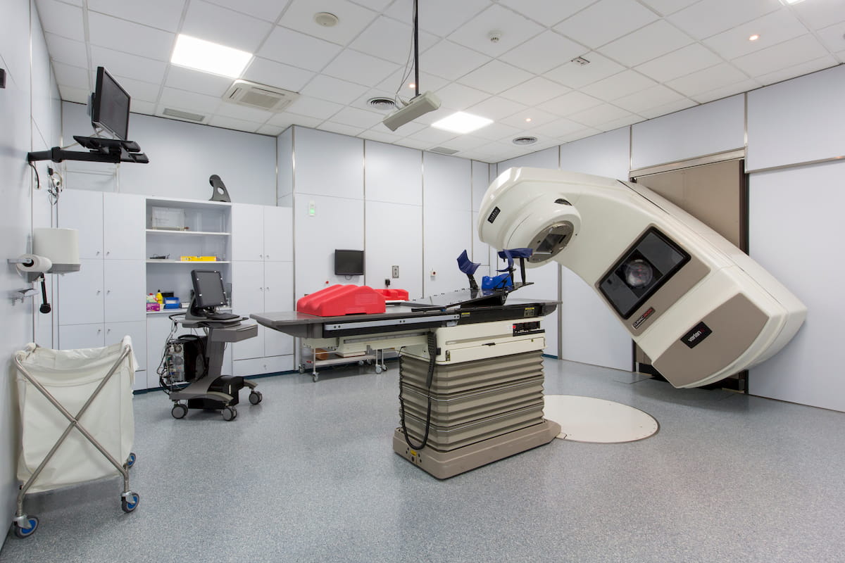
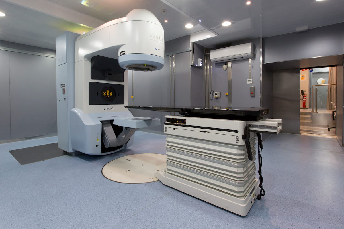
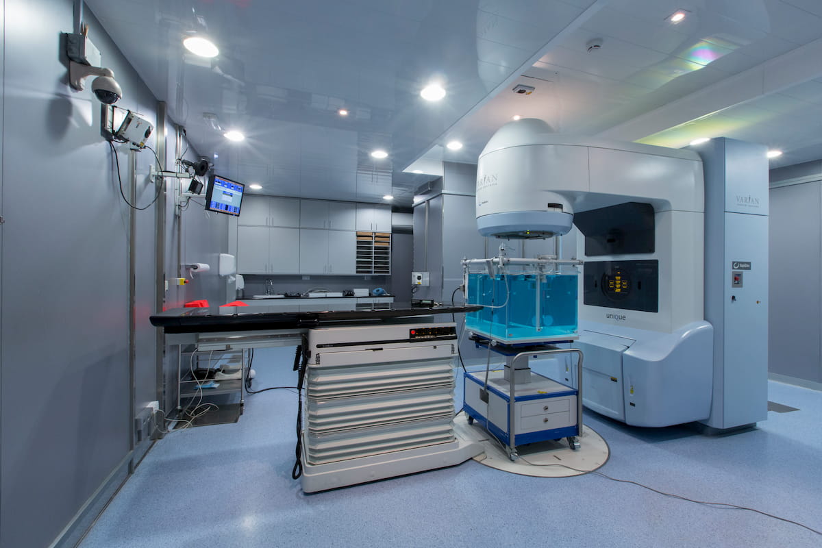
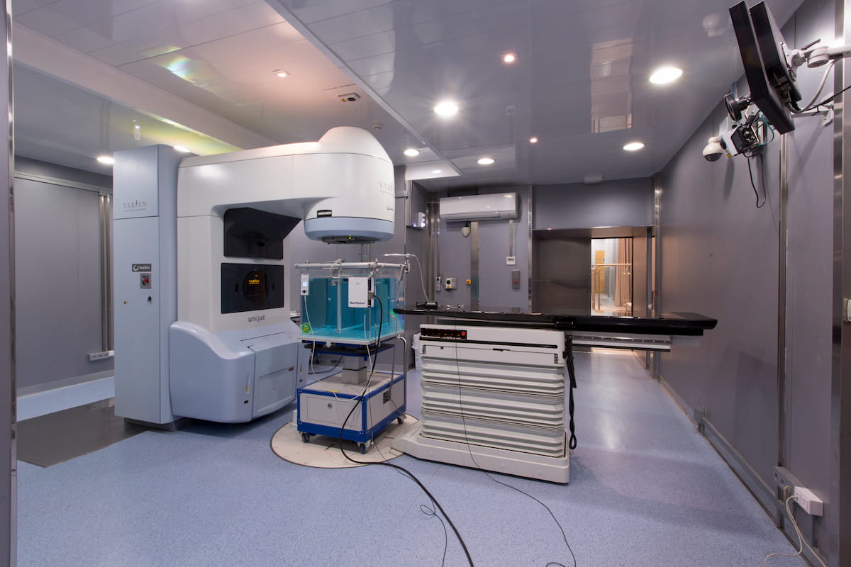
External radiation therapy
With the latest linear accelerators, the sessions are more effective and faster, allowing to apply high doses of radiation with better results in less time.

IGRT (Image-guided radiation therapy)
This type of radiotherapy allowed us ensuring that neighboring healthy structures that should not be irradiated remain outside the irradiation fields.

IMOR -
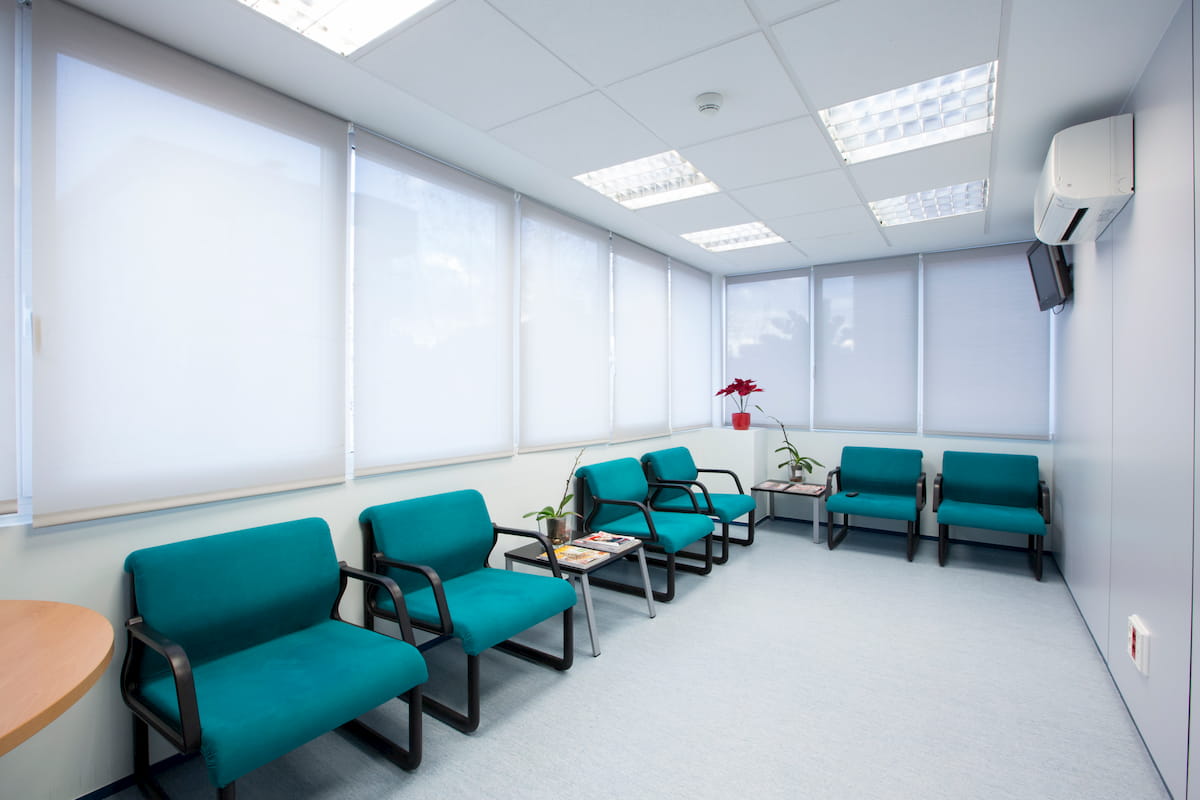
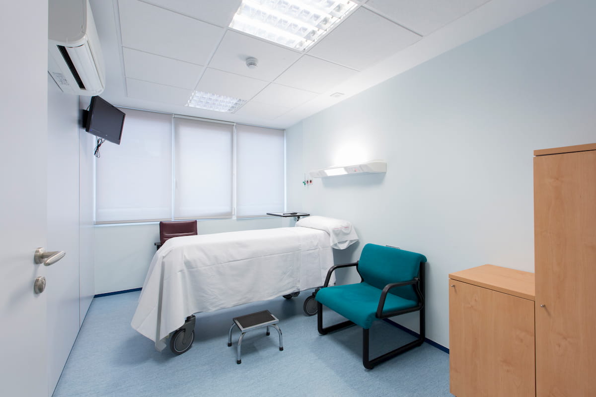
Oncological Hospital in Barcelona
The IMOR Institute, Medical Institute of Oncological Radiotherapy, is a oncological institute that offers various treatment modalities for cancer.
This foundation has been awarded by American Brachytherapy Society with the Judith Sitt Prize, considered the world’s highest award, for its outstanding contribution to the conservative treatment of prostate cancer and to the continuous improvement of the quality of life of the patients with prostate cancer.
What cases do we deal with?
In addition to ongoing research in the fight against cancer, we offer medical care, information about this disease, either about its treatment or prevention.
We can help you with any sign indicating the occurrence of a tumor (first stages) or in advanced stages of cancer and metastasis. We have oncologists and radiotherapists for the cases of prostate cancer, lung cancer, uterine cancer, bladder cancer, gastric cancer (stomach, esophagus, liver, pancreas …), breast cancer, brain tumors or bone cancer among others.
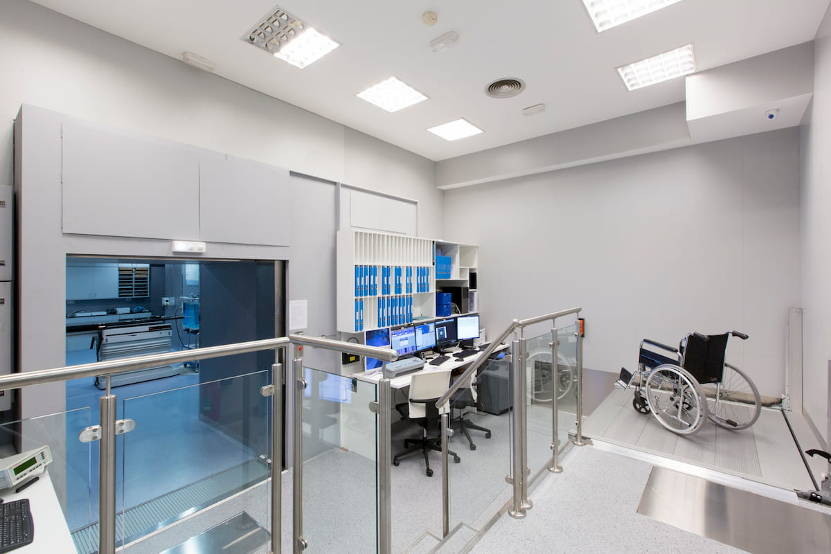
For treatments, we utilize the latest technology available in our field, the one that allows us to obtain the best results for the patient, both in terms of efficacy of the fight against cancer and in the health and well-being of the patient for the duration of the treatment and afterward, and this implies having techniques, systems and treatments that allow obtaining the least side effects.
In addition, some of these devices shorten the duration of the treatment that has been much longer. Now it is possible to carry out cancer treatment sessions in a few minutes.
Treatments for cancer
Any of these treatments do not have to be exclusive, and they can be complementary or parallel, even with chemotherapy, surgery or medication.

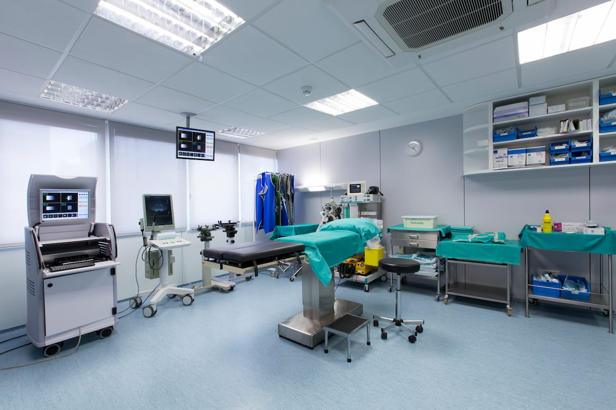
Intraoperative radiotherapy
This treatment consists of a radiotherapy session carried out in the surgical bed itself after the lumpectomy. It is usually used in tumors that appear in the breast or on the skin.
3D radiotherapy
It allows applying better treatments thanks to the use of patient images obtained from a CT tool in exactly the same position as that in which the treatment will be performed. It is an advantage for both the patient and the medical team.
4D Radiotherapy
It allows us to modulate radiation taking into account the movement of the tumors.
Brachytherapy
HDR brachytherapy treatment. Prostatic brachytherapy, ophthalmic brachytherapy and brachytherapy for other body areas with a tumor for which this type of treatment is optimal.
Other treatments
Extracranial radiotherapy treatment or SBRT (similar to radiosurgery but allowing to act directly on small tumors outside the head). There are also other treatments of stereotactic radiotherapy.
Publications
 The IMOR Foundation, Medical Institute of Onco-Radiotherapy has been awarded by the American Brachytherapy Society (ABS), with the Judith Sitt Prize, considered the highest award in the world, for its Outstanding Contribution to Conservative Cancer Treatment …
“Radiotherapy advances in lung cancer treatments”

Lung cancer is one of the most frequent in Spain. Contrary to what happens with some of the tumors more …
Get to know us better
Contact IMOR Institute
Are you looking for an oncology hospital with the best treatments for cancer in Barcelona? This website explains different ways to contact us for any question related to the prevention of the disease and all kinds of information about our cancer treatments. Of course, you can also contact us if you need medical attention urgently. We can help you.